Fig. 6.
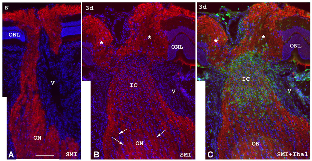
SMI312 (red) and Iba1 (green) immunolabeling of the ON in a control ON (A) and an rAION-induced ON three days after induction (B and C). Sections are counterstained with Hoechst for the cell nuclei. (A) The normal ON shows intact neurofilaments characterized by intense SMI312 immunostaining in the anterior, intrascleral and retroscleral portions of the ON. (B and C). In a rAION-induced ON, SMI312 staining is intense in the anterior and retroscleral ON (arrows in panel B), but there is significant disruption of labeled neurofilaments in the axons in the intrascleral and immediate retroscleral portion of the ON (IC, ischemic core in panel B), with heavy infiltration of Iba1(+) macrophage/microglia (green in panel C). This likely represents the ischemic infarct region. Significant disc edema (*) is also noted in the anterior portion of the ON at 3d post ischemia. The juxtapapillary ONL layer is displaced laterally from the ON edema caused by the infarct in the intrascleral and immediate retroscleral region. (ONL: outer nuclear layer; V: blood vessel.) Bar=50 µm. (For interpretation of the references to colour in this figure legend, the reader is referred to the web version of this article.)

