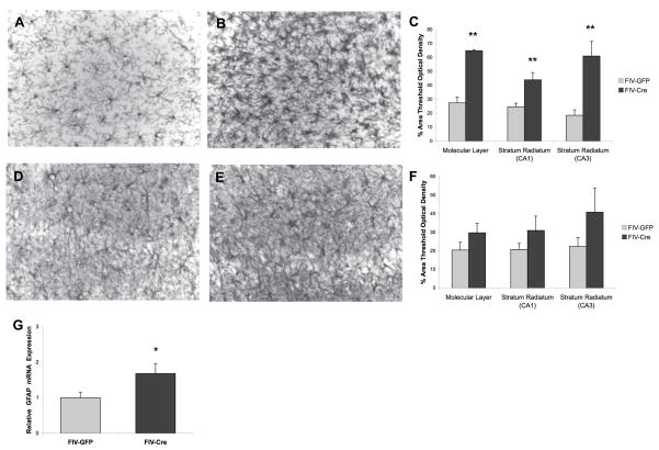
Figure 6.
Induction of hippocampal IL-1β leads to reactive gliosis. FIV-Cre-injected mice show evidence of microglial and astrocytic hypertrophy based on visual observation of Iba-1 and GFAP (B, E) immunoreactivity, respectively, relative to FIV-GFP-injected (A, D) mice. Analysis evaluating percent of area reaching threshold optical density showed increased immunoreactivity of Iba-1 (C) and GFAP (F) in FIV-Cre hippocampal regions. ** indicates p<0.01 relative to FIV-GFP animals; n=4 per experimental group. Bar in E = 10 μm (G) Real-time PCR showed elevated hippocampal GFAP mRNA associated with FIV-Cre injection. * indicates p<0.05 relative to FIV-GFP animals; n=9–12 per experimental group.

