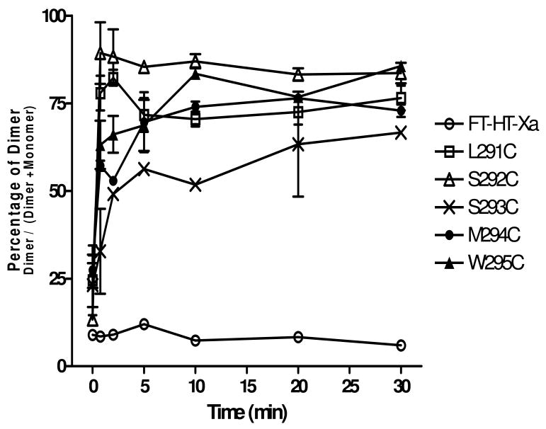
Figure 5.
Time course analysis of dimer formation at residues C-terminal to P290 of TM7. Total membranes were prepared from cells expressing each receptors and treated with Cu-P (Cu (II)-1, 10-phenanthroline) for 45 sec, 2 min, 5 min, 10 min, 20 min, and 30 min. The samples were analyzed by SDS-PAGE, following by immunoblotting with anti-FLAG antibody. The average percentage of dimer population from three independent experiments was calculated and plotted.

