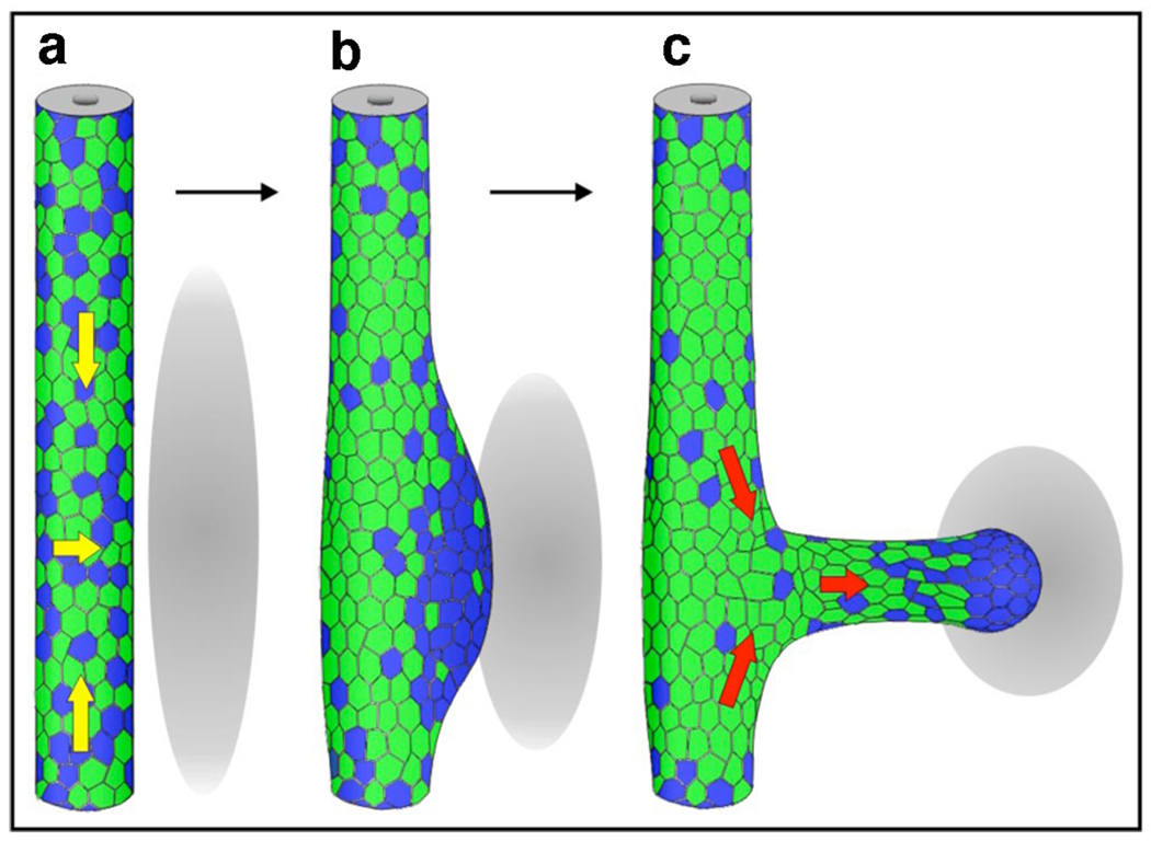
Figure 1. Cell movements and heterogeneous Ret signaling during ureteric bud formation.
Diagram illustrating rearrangement of Ret “positive” (blue) and Ret “negative” cells (green). Gray ovals represent the metanephric mesenchyme. Initially Ret “positive” cells are dispersed along the WD (A) and start to move (yellow arrows) to form the primary ureteric bud (UB) tip domain (B). As the UB grows out, these cells “lead” to form the distal tip, while Ret “negative” cells follow (C, red arrows). Reproduced with permission from [25]

