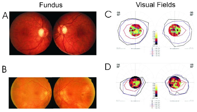
Figure 1.

Fundus photographs and visual field tests of 2 female patients with anti-transducin autoantibodies–Patient #5, age 73-years (A and C) and Patient #14, age 82-years (B and D): A: Mild temporal pallor of the disc. B: Normal fundus appearance. Bottom: Octopus 101 (Interzeag, Berne-Koniz, Switzerland) kinetic perimetry with overlying static fields showing mild (C) and moderate (D) constriction for all isopters tested with patchy, more severe loss of central retinal sensitivity on static testing.
