
Figure 4.
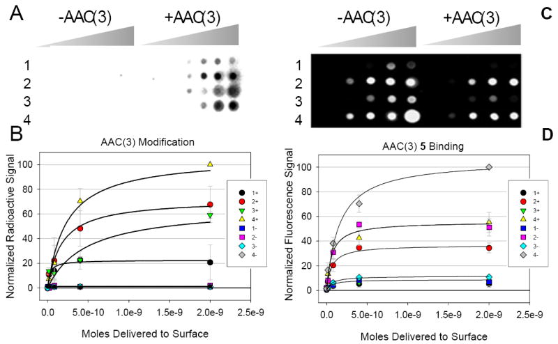
Modification of aminoglycosides by AAC(3) on a microarray and the subsequent effect on binding to an oligonucleotide mimic of the bacterial A-site, 5. A, a representative image of modification of aminoglycosides 1, 2, 3, and 4 (Figure 2) by AAC(3). The two halves of the microarray were spotted in the same manner. The left side was incubated with a cell lysate that does not contain AAC(3) and [14C]acetyl-CoA. As expected, there is no transfer of the radioactive tag to the aminoglycosides immobilized on the surface. The right side labeled “+AAC(3)” was incubated with AAC(3) and [14C]acetyl-CoA. B, Plots of the data from two microarray experiments represented in Panel A. 1+ is the data for compound 1 when incubated with a cell lysate containing AAC(3); 1- is the data for compound 1 when incubated with a cell lysate that does not contain AAC(3); 2+ is the data for compound 2 when incubated with AAC(3) while 2- was not incubated with AAC(3), etc. C, the microarray in panel A was probed fro binding 5 after 1, 2, 3, and 4 were modified by AAC(3). The left side of the microarray was incubated without AAC(3) while the right side was incubated with AAC(3). D, Plots of data for two microarray experiments represented in Panel C.