
Figure 5. Effect of PI3 kinase inhibition after onset of VCD-induced ovotoxicity.
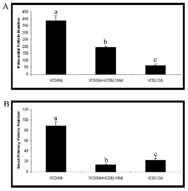
Ovaries from PND4 F344 rats were cultured with medium containing VCD (30 μM) for 6d. After the initial 6d of treatment, ovaries were removed from culture, or treated with VCD (30 μM) ± LY294002 (LY29; 20 μM) for an additional 6d. Following incubation, ovaries were collected and processed for histological evaluation as described in materials and methods. (A) Primordial and (B) small primary follicles were classified and counted. Values are mean ± SE total follicles counted/ovary, n=5; Different letters differ from one another within each group; P < 0.05.