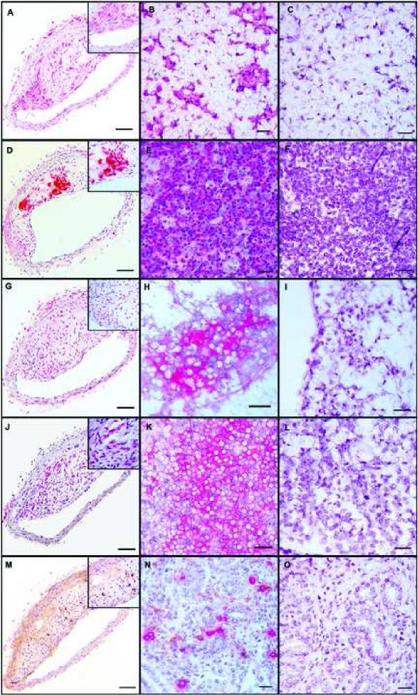
Figure 3.
Representative immunohistochemical staining of innominate arteries (left column), positive control tissue (center column) and negative control tissue (right column); 10X magnification, insets 40X magnification. A-C: anti-F4/80 monoclonal antibody staining for macrophages; positive and negative control tissue is lymph node. D-F: Anti-CD3 monoclonal antibody staining for CD3+ cells; positive and negative control tissue is thymus. G-I: Anti-CD4 polyclonal antibody staining for CD4+ cells; positive and negative control tissue is lymph node. J-L: Anti-CD8 monoclonal antibody staining for CD8+ cells; positive and negative control tissue is lymph node. M-O: Double immunostaining for NK cells (red) using anti-Ly49G2 monoclonal antibody and for dendritic cells (brown) using anti-CD11c monoclonal antibody; positive and negative control tissue is uterus.

