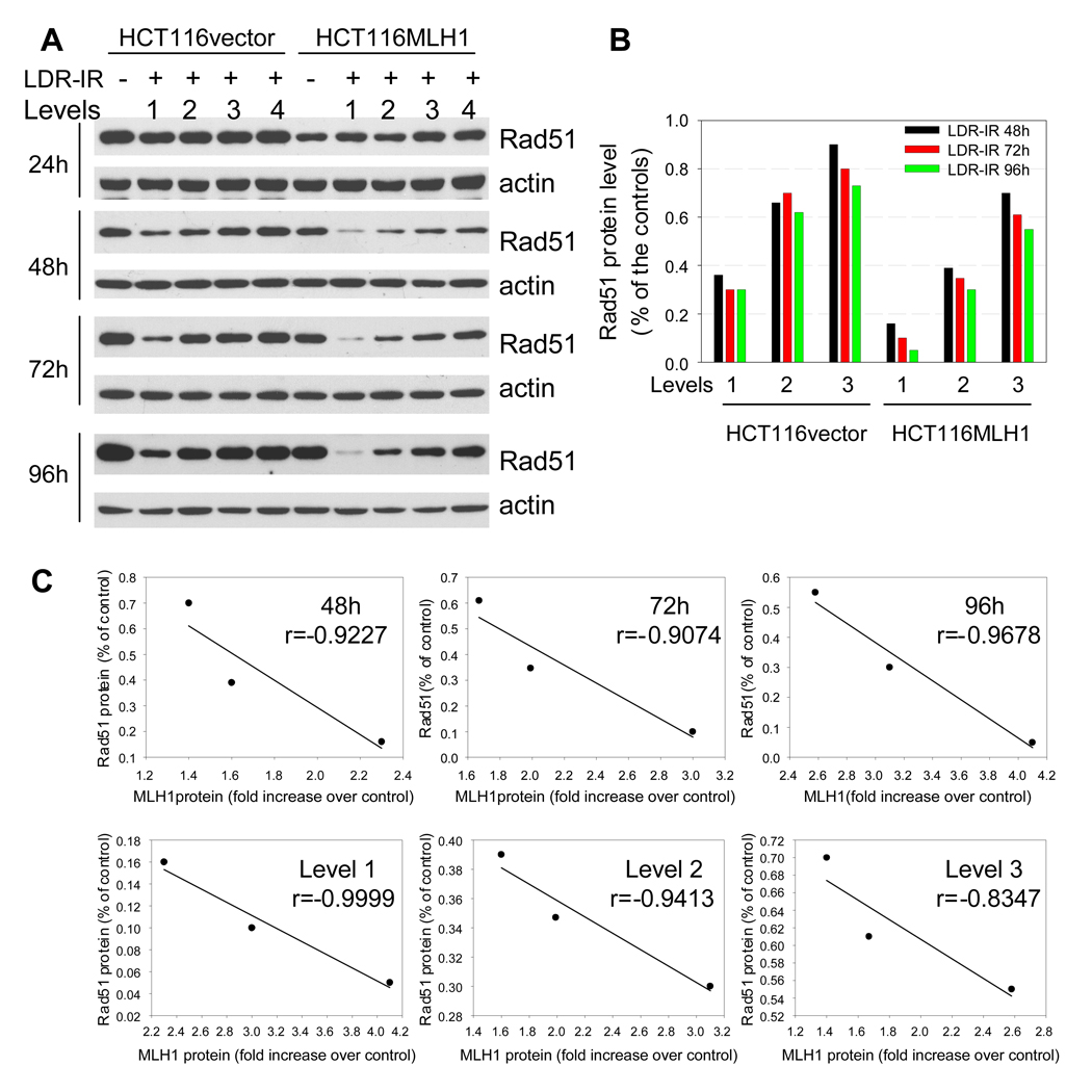
Figure 4.
Rad51 protein decreases during prolonged LDR-IR. A. Western blots demonstrate that Rad51 protein decreases progressively during prolonged LDR-IR with more significant reduction found in HCT116MLH1 (MLH1+) cells. B. Rad51 protein levels in Fig. 4A were quantified with software Image J. C. Linear regression test shows a strong correlation between MLH1 protein increase and Rad51 protein decrease in the MLH1+ cells. Correlation coefficients are calculated on data sets from Fig. 4B using Excel statistics. All Western blotting assays are repeated at least twice.

