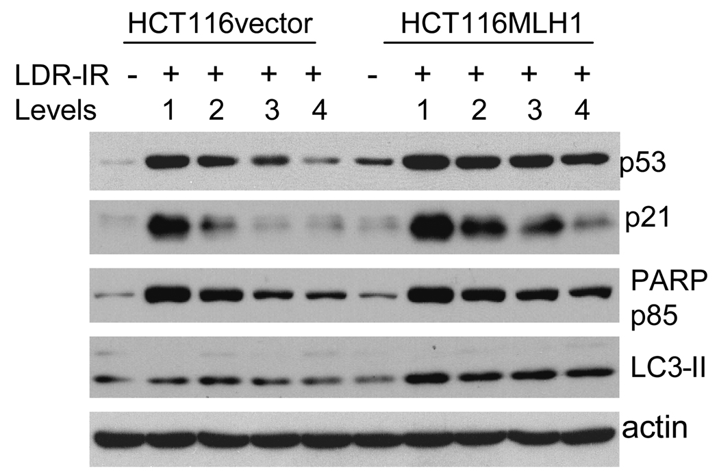
Figure 5.
Western blots demonstrate that levels of p53 and p21 proteins as well as cleaved PARP p85 fragment, are all elevated in both cell lines after a 72h exposure to prolonged LDR-IR but to a greater extent in the MLH1+ cells, while LC3-II is increased in the MLH1+ cells but not in the MLH1− cells. All Western blotting assays are repeated at least twice.

