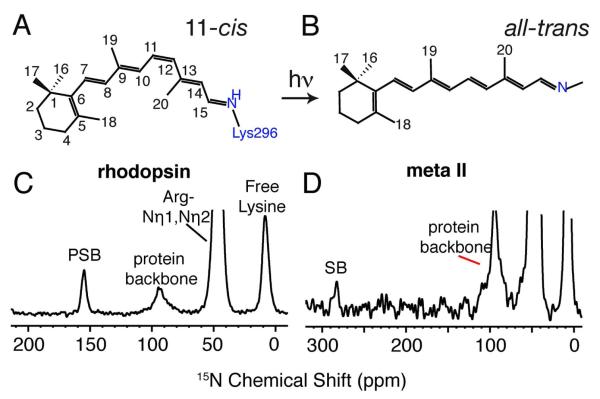
Figure 1.
Molecular structures of the 11-cis retinal PSB chromophore in rhodopsin (A) and the all-trans retinal unprotonated SB chromophore in Meta II (B). 15N CP MAS spectra of rhodopsin (C) and Meta II (D) labeled with 15Nε-lysine. The 15Nε-Lys296 is observed as a distinct narrow peak at 155.4 ppm in rhodopsin and shifts ~127 ppm downfield to 282.8 ppm in Meta II. The 15Nε resonances of the free lysines in rhodopsin are observed as a broad peak around 8.7 ppm.

