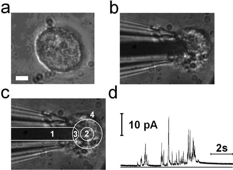
Figure 4.
(a) A peritoneal mast cell. Scale bar: 5 μm. (b) The mast cell of (a) on top of a single platinum electrode (PtE). (c) Different regions indicated by numbers. 1: SiO2 insulated part of Pt conductor, 2: PDL coated area, 3: exposed area forming the active PtE, 4: All the area outside the large circle including area 1 is covered with the SiO2 insulation layer. (d) The corresponding amperometric recording from the cell shown in B.

