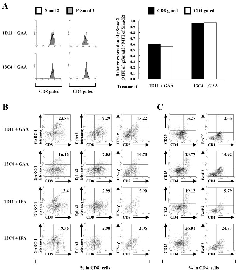
Figure 3. Systemic 1D11 promotes type-1 anti-GAA CTLs while reducing Treg cells in the i.c. glioma tissue.
Mice bearing orthotopic gliomas were treated with 1D11 and GAA vaccines as described previously. A, left panels, on day 25, BILs were isolated, and intracellular pSmad2 and Smad2 levels were evaluated in CD8- or CD4-gated populations by flow cytometry. Open and shaded histograms represent cells stained for Smad2 or pSmad2, respectively. Right panels, relative expression levels of pSmad2 in CD8-gated (black columns) or CD4-gated (white columns) population of the BILs calculated as relative mean fluorescence intensity (MFI) values of pSmad2 to those of Smad2 in corresponding cell populations. BILs were isolated from treated mice, and flow cytometric analyses were performed for (B) surface expression of CD8 and GARC-177-85 tetramer, EphA2671-679 tetramer or intracellular IFN-γ, and (C) surface expression of CD4 and CD25 or intracellular FoxP3 in lymphocyte-gated populations. For flow cytometric analyses of BILs (B and C), because of the small number of BILs obtained per mouse (∼4×10⁵ cells/mouse), BILs obtained from 5 mice per group were pooled, and evaluated for the relative number and phenotypes between groups. Numbers in each dot plot indicate the percentage of double-positive cells in (B) CD8-gated or (C) CD4-gated BILs. One of two representative experiments with similar results is shown.

