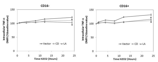
Figure 5. Oxidant exposure results in enhanced intracellular expression of TNF-α within monocyte subpopulations.
Human monocytes were stimulated with 100 mM H2O2 for up to 24 hours. Selected cells were pretreated with either 2 μM CD or 1 μM LA. Intracellular TNF-a expression was then examined with appropriate isotype control antibody by FACscan. Data are expressed as percentage change in mean fluorescence intensity (ΔMFI) compared to baseline conditions (set at 100%). Significant differences: * P <0.05 vs. time-matched control values based on 6 separately performed experiments.

