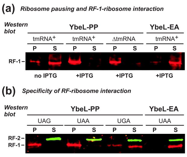
Figure 4. RF-1 interacts with the ribosome during translational pausing.
(a) Anti-RF-1 Western blot. Cell lysates were fractionated by sucrose density ultracentrifugation, and the pellet (P) and supernatant (S) fractions analyzed by Western blot. RF-1 was found primarily in the supernatant fraction of uninduced cells (no IPTG), and cells expressing YbeL-EA. In contrast, RF-1 partitioned to the pellet fraction in tmRNA+ and ΔtmRNA cells expressing YbeL-PP. (b) Anti-RF-1/RF-2 Western blot. Lysates from tmRNA+ cells expressing YbeL-PP from messages with UAG, UAA, and UGA stop codons were fractionated by ultracentrifugation, and analyzed by Western blot. RF-1 partitioned in the pellet fraction upon expression of the UAG and UAA constructs, but was found in the supernatant with the UGA message. RF-2 was found in the supernatant fraction for all constructs.

