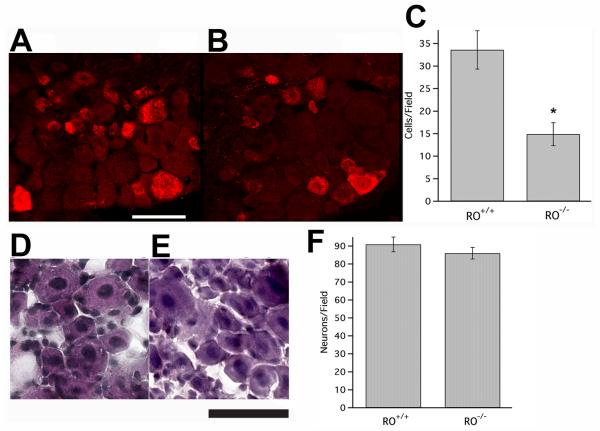
Figure 2. Decreased number of nociceptive neurons in adult (P60) PTPRO-/- mice.
(A,B) Confocal fluorescent micrographs of wt (A) and PTPRO-/- (B) P60 L4 DRG sections stained for CGRP to label nociceptive neurons. PTPRO-/- DRG demonstrate a >50% decrease in the number of nociceptive neurons per section (C). N=2 animals, 2 DRG, wt; N=2 animals, 4 DRG, PTPRO-/- .*, p < 0.02. (D, E) Bright field micrographs of wt (D) and PTPRO-/- (E) P60 L4 DRG sections stained for HE. There is no difference in the number of neurons per section between PTPRO-/- and wt (F, p>0.3). N=2 animals, 2 DRG, wt; N=3 animals, 5 DRG, PTPRO-/-. Scale bar, 50μm.

