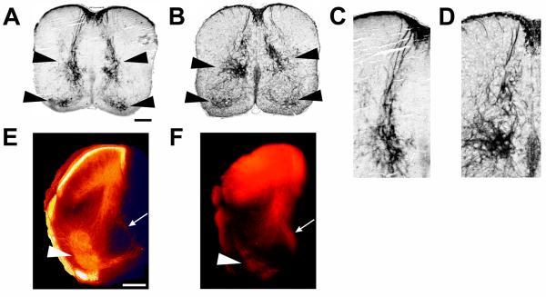
Figure 4. Aberrant proprioceptive projections in PTPRO-/- mice.
(A-D) P0 spinal cord sections stained with parvalbumin antibody to label proprioceptive projections in wt (A, C) and PTPRO-/- (B, D) mice. Arrowheads point to areas of aberrant axon projections. Disorganized and defasciculated axonal projections in PTPRO-/- mice are obvious in the magnified views (C, D). Additionally, the ratio of intermediate zone axonal projections to those in the ventral horn is higher in the PTPRO-/- mice. Both of these patterns were observed in all animals examined (n=7, wt; n=6, PTPRO-/-). (E, F) DiI labeled axon projections in P0 lumbar spinal cord. Fewer axons in PTPRO-/- mice reach their ventral horn motor neuron targets (arrowheads) compared to targets in the intermediate zone (arrows). Scale bars, 100μm.

