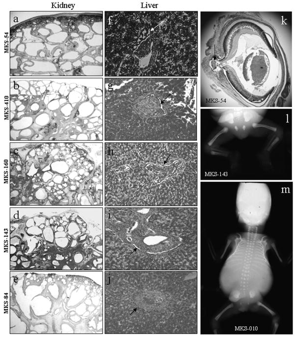
Figure 1. Phenotypic features of cases with CC2D2A mutations.
Histology pattern (hematoxylin-eosin stain) of kidney (a–e), liver (f–j) and eye (k) in CC2D2A-mutated cases. Kidney histology shows conserved corticomedullary organisation with little generation of mature glomerules. Cysts are found in the deep cortex and medulla, and are smaller at the periphery than in the center (a–e). Liver histology shows portal fibrosis with important and diffuse bile duct proliferation in all cases (arrows f–j). Sagittal section of the eye in case MKS-54 (k) shows an optic nerve cystic coloboma. X-rays show femoral bowing in case MKS-143 (l) and a bell-shaped thorax in case MKS-10 (m).

