
Figure 3. Brain MRI of a normal control (C), 6 month-old affected boy (JBS-006) and 13 year-old affected boy (JBS-008) carrying CC2D2A mutations.
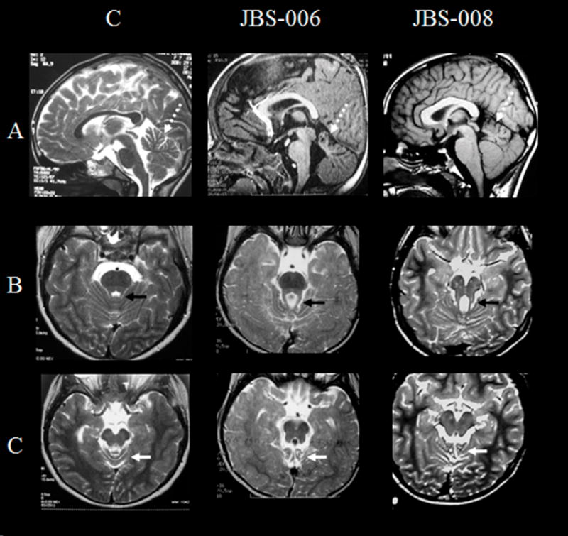
A: Sagittal images show in both affected patients a superior vermian dysgenesis. The middle and inferior segments of the vermis are hypoplastic or absent.
B: Axial T2-weighted FSE images demonstrate the abnormally thickened and elongated superior cerebellar peduncles and the molar tooth sign in all affected patients (black arrow)
C: Axial T2-weighted FSE images show superior vermian dysgenesis (white arrow) at the level of the cerebral peduncles in all affected patients.