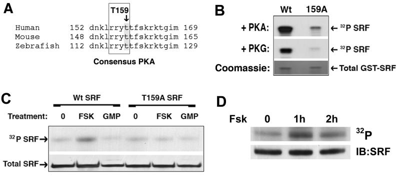
Figure 2. PKA phosphorylated SRF T159 in vitro and in vivo.
A) SRF sequence conservation near the consensus PKA/PKG phosphorylation site at T159 B) GST-SRF fusions (AA 1–203) containing Wt or T159A protein sequence were incubated with γ32P-ATP in the presence of active PKA or PKG for 15 min. Following removal of unincorporated label, samples were separated on an SDS-Page gel and exposed to film. C) Cos-7 cells expressing flag-tagged Wt and T159A SRF were incubated with 1mCi ortho 32P for 2h and then stimulated with 20μM forskolin or 100μM 8-pCPT-cGMP for 2h. SRF was then immunoprecipitated from RIPA lysates using anti-flag agarose. Following washing immunoprecipitants were run on an SDS-Page gel, transferred to nitrocellulose, and exposed to film. D) Endogenous SRF was immunoprecipitated from primary SMCs labeled with ortho 32P and then treated with FSK for the indicated times. Immunoprecipitants were run on an SDS-Page gel, transferred to nitrocellulose, and exposed to film.

