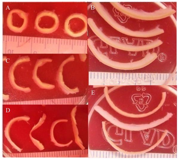
Figure 2.
Sample strips for material testing. A: arterial rings with small extracellular lipid pools; B: axial strips with fatty streaks; C: open-up sectors after radial cuts were made in the rings in (A); D: separated layers, adventitia (sectors with relatively round shape) and media (sectors with more irregular shape) of the open-up sectors; E: separated layers, adventitia (thicker strip) and media (thinner strips) of axial strips. Each small grid is 1mm width.

