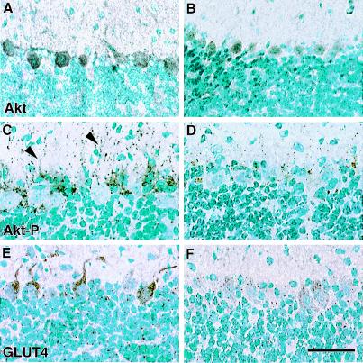
Figure 2.
Akt phosphorylation and GLUT4 localization in WT and Igf1−/− cerebellar cortex. Total Akt immunoreactivity is homogeneously distributed in Purkinje perikarya and processes in WT (A) and Igf1−/− (B) brains. Phospho-specific Akt immunoreactivity (Thr308), in contrast, is granular-appearing and is detected predominantly in Purkinje dendritic processes in the WT (C, arrowhead) and is scarcely detected in the Igf1−/− brain (D). GLUT 4 immunoreactivity is concentrated in Purkinje dendrites and the cytoplasm just around the dendrite's origin (E). GLUT4 is present in Purkinje perikarya in the Igf1−/− cerebellum, but is not preferentially localized in dendrites (F). (Bar = 100 μm.)

