
Figure 2.
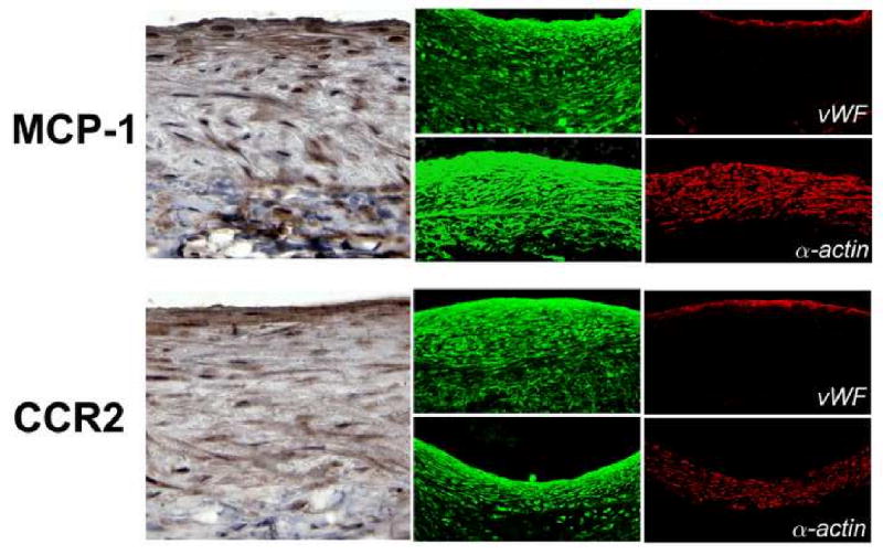
Distribution of MCP-1 and CCR2 in murine vein grafts 28 days following implantation. Von Willebrand Factor (vWF) and α-actin staining, for identification of endothelial and smooth muscle cells respectively, demonstrate a broad distribution of MCP-1 and CCR throughout the wall without cell-specific localization.