
Figure 4.
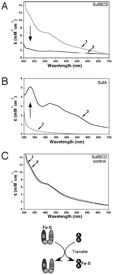
Fe-S cluster transfer from the SufBCD complex to SufA. Apo-SufA (300 μM) was incubated for 60 min with enough holo-SufBCD to provide a 3-4 fold molar excess of iron relative to the SufA concentration. SufA and SufBCD were then separated by anaerobic gel filtration and analyzed for Fe-S cluster content. Fe-S holo-SufBCD was prepared as described in Materials and Methods. Arrows indicate direction of change for spectra of holo-SufBCD or apo-SufA samples taken before transfer compared to spectra taken after transfer. (A) UV-visible absorption spectra of the SufBCD complex before (trace 1) and after (trace 2) the transfer reaction. (B) UV-visible absorption spectra of SufA before (trace 1) and after (trace 2) the transfer reaction. (C) Fe-S holo-SufBCD was incubated under the same conditions as the transfer reaction but without addition of apo-SufA. UV-visible absorption spectra of holo-SufBCD at time = 0 (trace 1) and time = 60 min (trace 2).