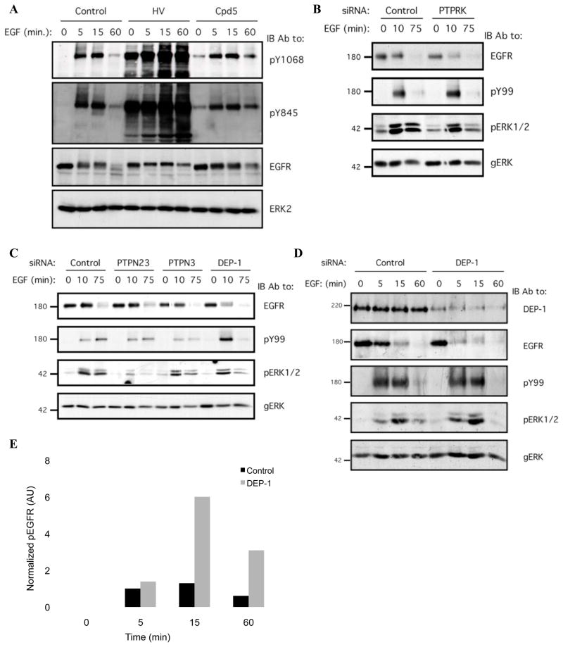
Figure 1. An Unbiased Genetic Screen Identifies PTPs Regulating EGF-mediated Receptor Phosphorylation and Degradation.
(A) HeLa cells were serum-starved for 16h, then treated with either a mixture of H2O2 (0.2mM) and sodium orthovanadate (1mM; for 15min; HV), Compound 5 (Cpd5; 20mM; for 30 min) or with ethanol (control; 30 min), and then stimulated with EGF (20ng/ml) for the indicated time intervals. Whole cell lysates were blotted with the indicated antibodies.
(B and C) Mixtures of siRNA oligonucleotides were transfected into HeLa cells, which were then incubated for 32h and serum-starved for 16h. The cells were then stimulated with EGF (20ng/ml) for the indicated intervals and lysed. Whole cell lysates were blotted with the indicated antibodies, including an anti-phosphotyrosine antibody (pY99), and antibodies to the active (pERK) and general forms of ERK (gERK).
(D) HeLa cells were transiently transfected with DEP-1 siRNA oligonucleotides or with control siRNA (each at 10nM), incubated for 32h, serum-starved for 16h and stimulated with EGF (20ng/ml) for the indicated time intervals. Whole cell lysates were immunoblotted with the indicated antibodies.
(E) HeLa cells were treated and processed as in D. EGFR phosphorylation was quantified using densitometric analysis and normalized to total EGFR level. One representative experiments (n=3) is shown.

