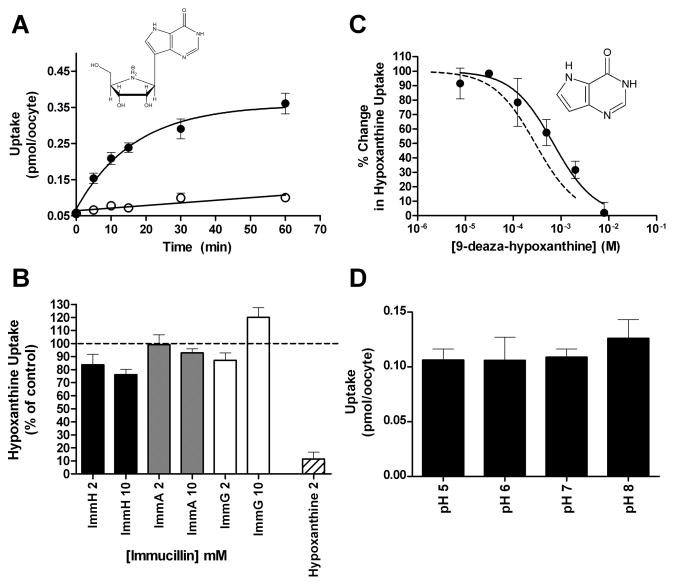
Fig. 7.
PfENT1 does not exhibit specificity for immucillin compounds. A. A time course for the uptake of 1.5 μM [5′-3H] Immucillin H by PfENT1 expressing (●) and DEPC water injected oocytes (○). The chemical structure of Immucillin H is shown. B. Competition of 2.5 mM or 10 mM Immucillin H (black bars), Immucillin A (grey bars), and Immucillin G (white bars) with 10 minutes of uptake of 100 nM [2,8-3H]hypoxanthine. Competition of 2.5 mM hypoxanthine (hashed bar) with 10 minutes of uptake of 100 nM [2,8-3H]hypoxanthine is shown. C. The structure of 9-deaza-hypoxanthine is shown. A range of 9-deaza-hypoxanthine concentrations (●) competes with 10 minutes of uptake of 100 nM [2,8-3H]hypoxanthine. Competition of a range of hypoxanthine concentrations with 10 minutes of uptake of 100 nM [2,8-3H]hypoxanthine was shown in Fig. 3A and is reproduced in this figure (dashed line) for purposes of comparison. D. PfNT1 mediated uptake of 1.5 μM [5′-3H]Immucillin H over a range of buffer pH values. For each pH, 10 minutes of ImmH uptake by PfNT1 expressing oocytes was measured and uptake by DEPC water injected oocytes is subtracted to account for background.

