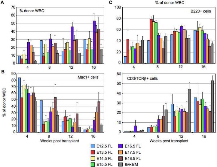
Figure 2.
LT-HSCs differ in their engraftment levels and lineage potential throughout fetal development and early adulthood reminiscent of long-term stem cell aging reconstitution kinetics. (A): Quantitative (mean ± s.e.m.) and kinetic analysis of the percentage of donor-derived total WBC in the peripheral blood of lethally irradiated CD45.1+ mice transplanted with 25 KTLS cells from donor CD45.2+ animals (E12.5, E13.5, E14.5, E15.5, E16.5, E17.5 and E18.5 FL, and 8 week BM) and 3×10⁵ recipient-type competitor adult whole BM (WBM). Donor cell reconstitution was assayed via readout in lysed, TER119- peripheral blood cells of recipients up to 16 weeks post transplantation. (B-C): Quantitative (mean ± s.e.m.) and kinetic analysis of the lineage potential of differently-aged KTLS cells showing the percentage of Mac1+ (myeloid, M lineage) (B), B220+ (B lineage) and CD3/TCRβ+ (T lineage) (C) cells within the donor-derived WBC compartment. The number of animals for which each of these statistics was calculated is shown in Table S1.

