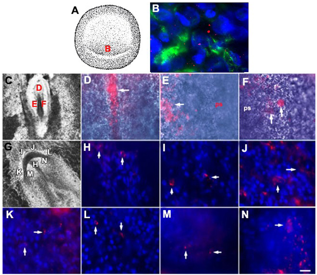
Figure 1. Identifying and tracking MyoD+/G8+ cells from the stage 2 epiblast into tissues of stages 5 and 7 embryos.
Whole stage 2 embryos were double labeled with the G8 MAb and fluorescein conjugated secondary antibodies (green) and Cy3 dendrimers to MyoD mRNA (red) (B). Nuclei were counterstained with Hoechst dye (blue). The area indicated in the drawing of the stage 2 embryo (A) is shown at high magnification in B. MyoD+ epiblast cells of the stage 2 embryo express the G8 antigen and MyoD mRNA (B). Whole stage 2 embryos were labeled with the G8 MAb and rhodamine conjugated secondary antibodies and incubated until they reached stages 5 (C–F) or 7 (G–N). Stage 7 embryos were counterstained with Hoechst dye. The areas designated with a letter in the DIC images of the embryos (C and G) are shown at high magnification in the remaining photomicrographs. Arrows indicate the locations of some of the fluorescent G8+ epiblast cells in whole embryos. By stage 5, G8+ epiblast cells had been incorporated into the head process (D). The epiblast on either side of the anterior portion of the primitive streak also contained G8+ epiblast cells (arrows in E and F). By stage 7, G8+ epiblast cells were found in the prechordal mesoderm (H), the anterior (I and J) and anterior/lateral preplacode regions of the ectoderm (K and L), and the anterior/lateral neural plate (M and N). Scale bar: 135 µm in C and G; 9 µm in D–F and H–N.

