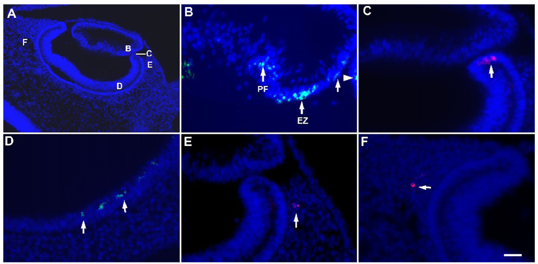
Figure 2. Distribution of G8+ epiblast cells in the eyes of stages 17 and 18 embryos.
Stage 2 embryos were labeled with the G8 MAb and a rhodamine (red) or fluorescein (green) conjugated secondary antibody, incubated to stages 17 or 18, fixed and sectioned. Nuclei were counterstained with Hoechst dye (blue). A representative section through the stage 17 eye is shown in panel A. The areas indicated by the letters in panel A are shown at high magnification in B–F. G8+ epiblast cells were present in the equatorial zone (EZ) and primary fiber region (PF) of the lens (arrows in B) and the anterior margin of the optic cup (arrowhead in B, arrow in C). G8+ epiblast cells also were present in the retinal layer of the posterior optic cup (D) and periocular mesenchyme (E and F). Scale bar: 27 µm in A; 13 µm in B–F.

