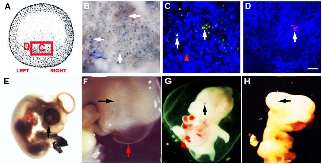
Figure 4. Lysis of MyoD+ cells in the epiblast and its effects on eye morphogenesis.
MyoD+ cells in the stage 2 epiblast were labeled with the G8 MAb and lysed with complement. The area in the box in the drawing of the stage 2 embryo in panel A is shown in panel B. The letters correspond to the areas shown at higher magnification in C and D. The uptake of trypan blue reveals the presence of lysed cells (B). A fluorescein conjugated secondary antibody was applied following treatment with complement (green). After incubating the embryos for three hours, the G8 MAb was reapplied and tagged with a secondary antibody conjugated with rhodamine (red). The overlap of red and green appears yellow-white in merged images. Most labeled cells were tagged with both fluorochromes (arrows in C). Nuclei appeared fragmented (arrowhead in C). Cells that expressed the G8 antigen within three hours after ablation were labeled with only rhodamine (D). These cells were concentrated on the left side of the epiblast a field away from the cells shown in panel C. The right (E) and left eyes were normal in control embryos treated with the E12 MAb and complement. One ablated embryo had an absence of visible eye tissue on the right (black arrow) and a normal eye on the left (red arrow) (F). Other embryos exhibited micropthalmia (G) or a lack of pigmentation (H). Scale bar: 56 µm in B; 9 µm in C–D.

