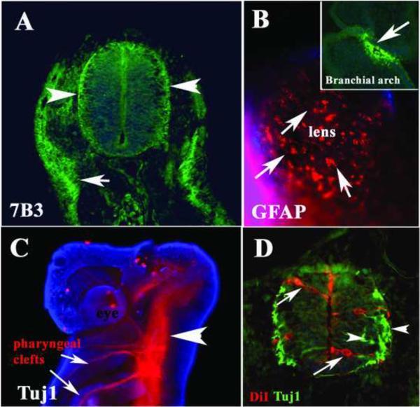
Figure 1. Neural markers expressed in the Stage 25 shark embryos.
Immunofluorescence of st.25 shark embryo for 7B3/transitin (A) GFAP (B) and Tuj1 (C, D). A Thin section through one embryo at the mid-trunk level shows positive 7B3 cells in the neural tube, especially on the ventricular and laminar side of the neural tube (arrowheads) and developing lateral line underneath the ectoderm (arrow). B Ectoderm around the eye shows cells positive for GFAP (arrows). Head (C) and trunk (D) immunostaining with the tubulin-III neuron specific antibody Tuj1 showed positive neural tube and axonal projections into the branchial arches (C). Cell nuclei were stained blue with DAPI.

