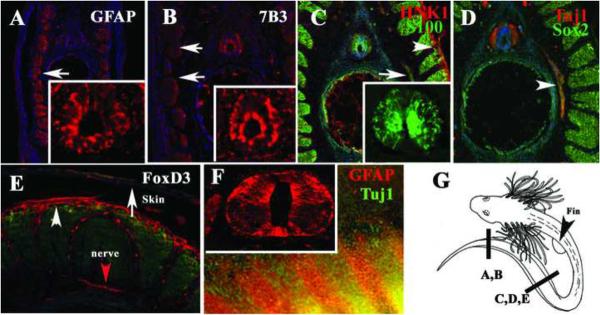
Figure 4. Glial and neural crest markers expressed in the Stage 32 shark embryos.
Sections through a st.32 embryo at the tail most end for GFAP (A), 7B3 (B), S100 (C), Tuj1 (D) and FoxD3 (E) labeled a specific group of cells. Radial glia and myoblasts were strongly stained with GFAP (insert for neural tube magnification in A and F). 7B3 labeled radial cells in the neural tube (insert in B) and to a lesser extent in the medial myoblasts (arrows). C S100 (green) also labeled radial cells; insert shows a magnification of the neural tube, while HNK-1 labeled skin (arrowhead). D Tuj1 (red) and Sox2 (green) weakly overlap and double labeled peripheral nerve (arrowhead in D). E FoxD3 was present in peripheral nerve (red arrow) as well as in dermal cells (arrowhead in E). GFAP labeled a group of cells in the fin (red arrows in F) while Tuj1 presented a banded staining in the fin (green). G shows a cartoon of a stage 32 embryo with line indicating the area for each figure. Cell nuclei were stained blue with DAPI.

