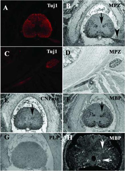
Figure 7. Myelin markers expressed in the trunk of pre-hatching shark embryo CNS.
Spinal cord sections were immunostained for Tuj1 and MPZ (A–D), CNPase (E), MBP (F, H) and PLP (G). Spinal cord and nerves had abundant neuronal processes as attested by Tuj1 and myelinated fiber are beginning to delineate the butterfly shape of mammalian spinal cords observed in osteichthyans (B, F and to lesser extent E). At the tail-most end, MBP labeled fewer axons along the same locations as in more rostral regions (H). PLP did not give positive staining on these sections (G). Arrows point to myelinated tracts.

