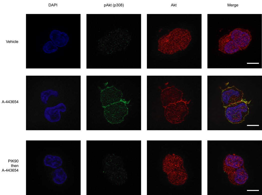
Figure 5. The Akt inhibitor A-443654 induces Akt membrane localization.
After treatment of HEK293 cells with various conditions described below, cells were fixed and stained with rabbit anti-Akt or mouse anti-pAkt (p308) followed by Alexa 488-conjugated goat anti-rabbit and Alexa 568-conjugated goat anti-mouse, and examined by fluorescence microscopy. Cells were treated with vehicle (DMSO) for 15min (top panel), treated with 2.5 µM A-443654 for 15 min (middle panel), or treated with 2.5 µM PIK90 for 10 min prior to the addition of 2.5 µM A-443654 for further 15 min (bottom panel). Scale bar 10 µm.

