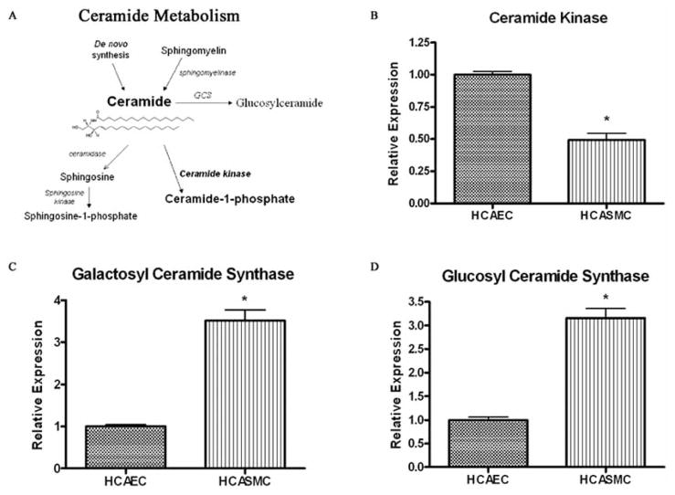
Fig. 3. Human coronary artery endothelial cells have an increased capacity to metabolize ceramide into phosphorylated, but not glycosylated, metabolites.
(A) Primary ceramide metabolic pathways. The dynamic flux of ceramide metabolites regulates the switch between apoptosis/growth arrest and proliferation. Anti-apoptotic/proliferative phosphorylated metabolites are formed via ceramide kinase or sphingosine kinase. The galactosyl- and glucosylceramide synthases (GCS) metabolize ceramide into less bioactive glycosylated ceramide species. (B-D) Gene expression of sphingolipid metabolic enzymes ceramide kinase (B), galactosylceramide synthase (C), and glucosylceramide synthase (D), in human coronary artery endothelial cells (HCAEC) and smooth muscle cells (HCASMC) was measured by real time RT-PCR. Enzyme mRNA expression levels are all relative to their respective level in vehicle treated HCAECs, (mean ± SEM, n ≥ 5); * Denotes significance compared to vehicle treated HCAECs.

