
Figure 3.
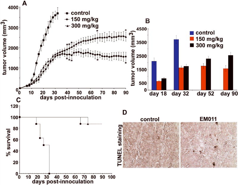
Orally fed EM011 inhibits tumor progression in vivo. Palpable tumors (~100 mm3) were established 6-8 days after injecting 106 B16LS9 melanoma cells (in 0.2 ml of PBS) subcutaneously in mice with a syngeneic background. A shows progression profile of tumor growth in control vehicle-treated mice compared with 150 and 300 mg/kg EM011 treatment. B depicts tumor volume measurements on days 18, 32, 52 and 90 post-inoculation. On day 32, control vehicle-treated mice were euthanized because of overgrown tumors, in compliance with the experimental IACUC protocol. Interestingly, treatment at 150 mg/kg showed better antitumor outcomes compared to 300 mg/kg. C shows Kaplan-Meier analysis of the therapeutic effect of two dose regimes (150 and 300 mg/kg) of EM011. (*, P< 0.05, A-C). D. Immunohistochemical TUNEL-staining of paraffin-embedded tumor sections from mice treated with vehicle (left panel) or EM011 (right panel) for 32 days (end point of control vehicle-treated animals). Right panel shows TUNEL-positive cells (seen as apoptotic brown nuclei) compared to left panel (vehicle-treated control).