
Figure 1. Localization of exogenous Sam68 into SG upon oxidative stress.

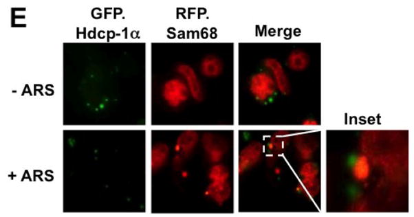
293T cells were transfected with RFP-Sam68 and GFP.TIA-1 (A), GFP.G3BP (B), GFP.hnRNP A1 (C), GFP.PABP (D), GFP.hdcp1α (E). Cells were cultured for 48 hr and then treated with 0.5 mM ARS (+ ARS) or without ARS (− ARS) for 1 hr prior to fixation and microscopic imaging. The images were representative of each transfection from at least three independent experiments. Co-localization of each marker with exogenous Sam68 was shown in the column marked as “Merge”.