
Figure 2. IL-1β is sufficient and required to activate Wnt signaling.
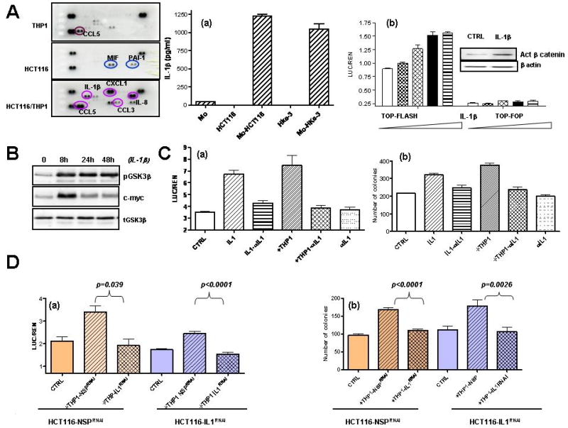
A: HCT116 and THP1 cells were cultured alone or together as indicated for 48 hours, and the expression of soluble mediators was determined by Human cytokine array (R&D Systems). (a): The amount of IL-1β was determined by ELISA in supernatants of HCT116 and Hke-3 cells, normal human monocytes (Mo) and in co-cultures. (b): HCT116 cells were transfected with TOP-FLASH or TOP-FOP reporter genes and were treated with increasing concentrations of IL-1β (0.5-10 ng/ml). Inset: The amount of active β-catenin in IL-1β treated cells. B:The levels of pGSK3 (Ser9), total GSK3 and c-myc in control and IL-1β treated HCT116 cells. C: (a) HCT116 cells were transfected with TOP-FLASH reporter gene and were treated as indicated. (b) HCT116 cells were treated as indicated and the clonogenic assay was performed as described in Material and methods. D: HCT116 were transfected with NSP (nontargeting) siRNA or siRNA specific for IL-1β. They were cultured with nontransfected THP1 cells or THP1 cells transfected with NSP siRNA or siRNA specific for IL-1β. Wnt signaling (a) and clonogenic assay were (b) were performed as indicated.