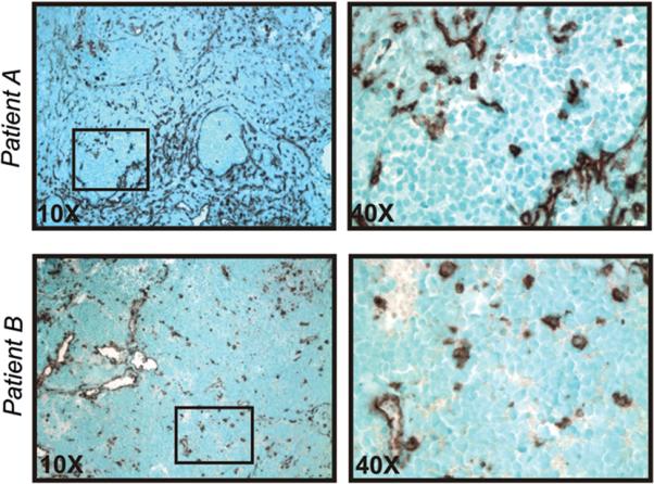
Figure 1.

MHC class I expression in NB tumors. Paraffin sections of high-risk NB tumors at diagnosis were stained with anti-MHC class I mAb. Results for Patient A (stroma-rich tumor) and Patient B (stroma-poor tumor) are shown. Black boxes in 10x panels (left) indicate areas of MHC class I-negative tumor nests (magnified in right panels); certain stromal elements, blood vessels, and tumor-associated macrophages are MHC class I-positive. Similar results were obtained for a total of 26 patients with HRNBL.
