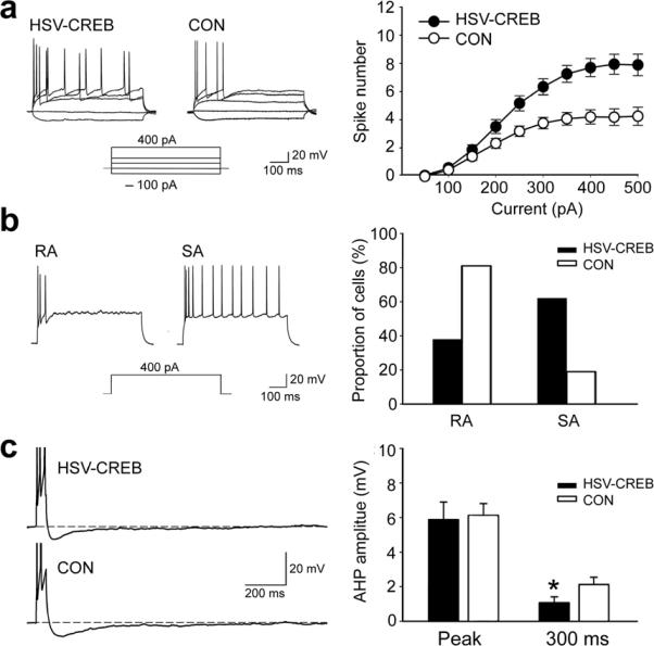
Fig. 5.

CREB increases neuronal excitability in transfected LA neurons. (a) Left: traces of sample recordings showing more APs in response to increasing current injections (−100, 0, 100, 200 and 400 pA; 600 ms) in a HSV-CREB neuron than in a neighboring non-transfected neurons (CON). Right: a plot of the number of evoked spikes as a function of injected current shows significantly more APs in HSV-CREB neurons (n = 58, from 8 mice) than in controls (n = 64, from 12 mice from the other three groups, Repeated ANOVA, P < 0.05). (b) HSV-CREB changed the firing properties of transfected LA pyramidal neurons. Left: sample voltage traces showing two LA neurons with distinct firing properties (RA and SA)24. Rapidly adapting (RA) cells fired only 1–5 spikes at the onset of the current injection (600 ms, 400 pA) and remained silent for the rest of the current pulses. Slowly adapting (SA) cells fire more than 6 spikes. Right: distribution of firing properties of HSV-CREB neurons and control LA neurons. (c) HSV-CREB neurons show reduced post-burst AHP compared to control neurons. Left: sample post-burst AHP traces from a HSV-CREB neuron and a neighboring control neuron. Right: histogram showing AHP difference between HSV-CREB neurons (n = 58, from 8 mice) and the control neurons (n = 64, from 12 mice from the other three groups) as measured at peak and 300 ms after the end of current step. Unpaired t test, *P < 0.05 as indicated.
