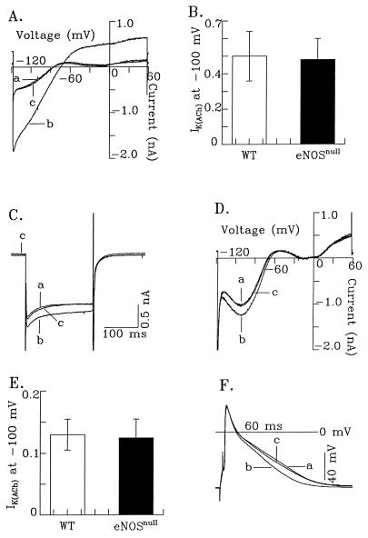
Figure 4.
Targeted disruption of eNOS does not affect muscarinic activation of acetylcholine-sensitive K+ current (IK(ACh)). (A) The activation of IK(ACh) in an eNOSnull atrial myocyte is illustrated at a holding potential of −40 mV and after a ramp pulse from −150 to +60 mV, applied to the cell at 0.1V/s. ICa-L was blocked by 0.4 mM Cd2+. The fast inward sodium current also was blocked largely by Cd2+. (B) A comparison of IK(ACh) from eNOSnull (n = 5) and WT (n = 7) atrial myocytes. The amplitude of IK(ACh) was determined as the difference current level at −100 mV. (C) The activation of IK(ACh) in an eNOSnull ventricular myocyte. A hyperpolarization pulse from a holding potential of −40 mV to −100 mV was applied to the cell, and traces a, b, and c were recorded under control conditions, with CCh (1 μM) application, and after washout of CCh, respectively. (D) The current–voltage relationship of IK(ACh) in an eNOSnull ventricular myocyte. The ramp pulse was identical to that described in A. ICa-L and sodium current were blocked by Cd2+ (0.4 mM). Traces a, b, and c were recorded under control conditions, with CCh (1 μM), and with atropine (1 μM) in the continuous presence of CCh. (E) A comparison of IK(ACh) (at −100 mV) from both eNOSnull (n = 8) and WT (n = 7) ventricular myocytes. (F) Activation of IK(ACh) in an eNOSnull ventricular myocyte abbreviates action potential duration. Traces a, b, and c were recorded under control conditions (a), during CCh (1 μM) application (b), and with atropine (1 μM) in the continuous presence of CCh (c).

