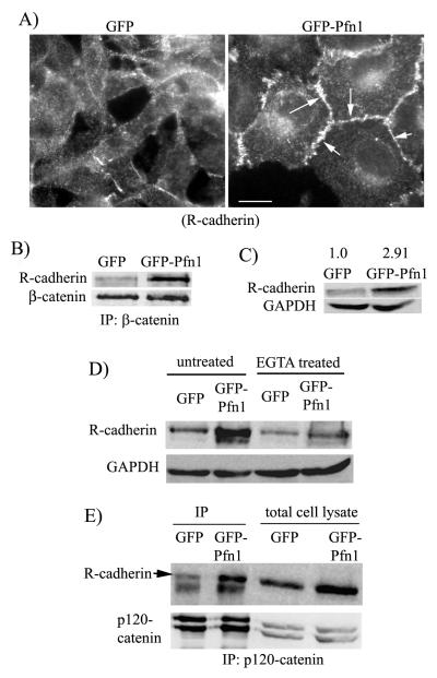
Figure 4. Pfn1 overexpression causes junctional accumulation of R-cadherin in MDA-231 cells.
A) R-cadherin immunostaining of GFP and GFP-Pfn1 expressers (arrows show junctional localization; scale bar — 20 μm). B) Co-immunoprecipitation analyses of R-cadherin/β-catenin complex formation in GFP and GFP-Pfn1 expressers. C) R-cadherin immunblot of total cell lysates shows ~2.9 fold increase in R-cadherin expression in Pfn1 overexpressing cells (the numbers are based on relative densitometric analyses of R-cadherin and GAPDH (loading control) bands averaged from 4 independent experiments). D) Relative R-cadherin levels between GFP and GFP-Pfn1 expressers either untreated or following 3 hours of 2 mM EGTA treatment (GAPDH blot serves as the loading control). E) P120-catenin and R-cadherin immunoblots of total cell lysate and p120-catenin immunoprecipitates (IP) from the lysates of GFP and GFP-Pfn1 expressers.

