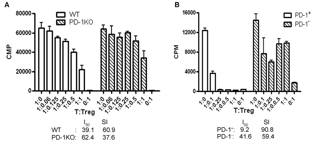
Fig. 4.
PD-1 expression was related to the suppressive activity of CD4+FoxP3+ Treg cells. A: PD-1 deficiency impaired the suppressive function of Treg cells from PD-1KO mice. B: CD4+PD-1+GFP+ Treg cells had greater suppression than CD4+PD-1−GFP+ Treg cells from FoxP3-GFP “knock-in” mice. CD4+FoxP3+ (in A) or CD4+PD-1+GFP+ and CD4+PD-1−GFP+ (in B) cells were sorted from splenocytes of immunized PD-1KO and WT B6 mice (in A) or FoxP3-GFP “knock-in” mice (in B). The suppressive activities of CD4+FoxP3+ cells from PD-1KO vs. WT mice or CD4+PD-1+GFP+ vs. CD4+PD-1−GFP+ from FoxP3-GFP “knock-in” mice were compared ex vivo by using the T-cell suppression assay. CD4+FoxP3− (in A) or CD4+GFP− (in B) cells from the same batch of mice were used as responder cells and T-cell-depleted splenocytes from naïve WT mice were used as APCs. The experiment was repeated twice with five mice per group in each experiment.

