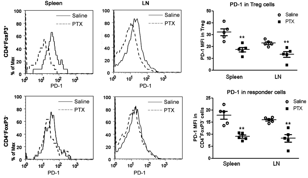
Fig. 6.
Administration of PTX reduced PD-1 expression in FoxP3+ and FoxP3− CD4 T cells. Six days after immunization with mMOG-35–55 peptide/CFA in the presence or absence of PTX, WT mice were euthanized, and the PD-1 expression levels in CD4+FoxP3+ and CD4+FoxP3− cells in both splenocytes and LN cells were analyzed. **P < 0.01 as measured by Student’s t-test (n = 5). The experiment was repeated twice.

