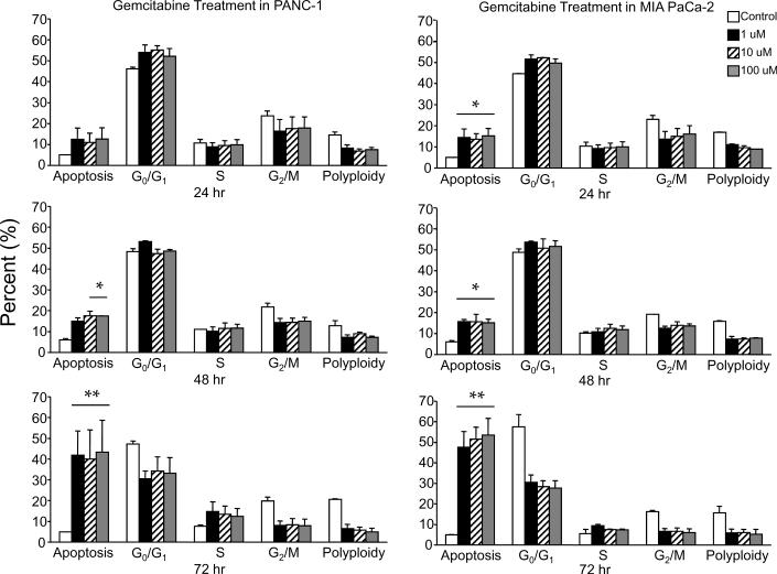
Figure 2.
Gemcitabine treatment of PANC-1 and MIA PaCa-2 cell lines. (A) Cells were treated using 0 μM,1 μM, 10 μM, and 100 μM gemcitabine after which they were harvested and analyzed for DNA content by propidium iodide staining and flow cytometry at 24 hr, 48 hr, and 72 hr. Percentages of apoptotic cells with hypodiploid (sub-G1) DNA content as well as those in G0/G1, S, G2/M and polyploid are indicated per each condition tested. Data are the mean ± SD of three independent experiments (*p<0.01, **p<0.001). (B) Detergent-solubilized extracts of pancreatic cells treated with gemcitabine were analyzed at the indicated time intervals for reactivity with antibodies for survivin and GAPDH (loading control), by Western blotting. Molecular-weight (Mr) markers in kilodaltons are shown on the left.


