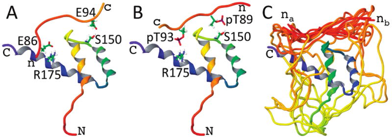
Figure 4.

Example model structures of (A) the AD peptide and (B) the phospho-AD peptide docked to the NKX3.1 HD. The HD ribbons are shaded from red at the N-terminus to blue at the C-terminus. These two examples highlight possible hydrogen-bonded contacts between either glutamate or phosphothreonine side chains of the peptides and the Ser150 and Arg175 side chains of the HD. (C) The reconstructed linker residues between the AD and HD are shown for an ensemble of docked peptide model structures. The linker can accommodate the peptide docked with either the peptide N-terminus closer to Arg175 (nA) or closer to Ser150 (nB).
