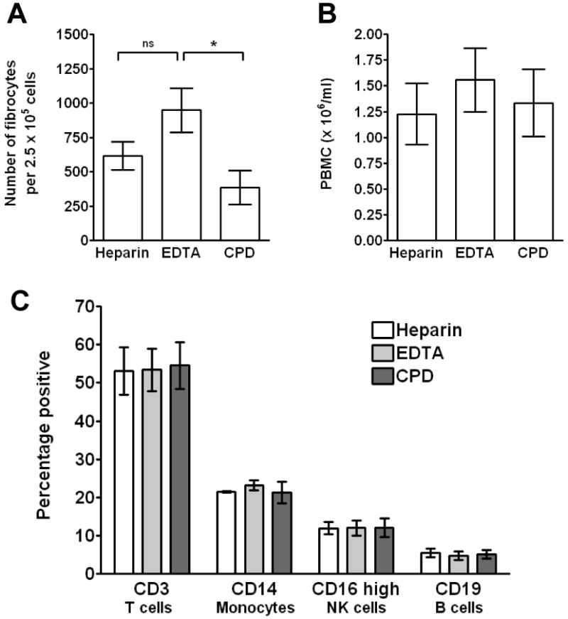
Figure 1. Effect of different anti-coagulants on fibrocyte differentiation.

A) PBMC separated from heparin, EDTA, or CPD-treated blood were cultured in RPMI-based serum-free medium for 5 days at 2.5 × 10⁵ cells per ml. Cells were then air-dried, fixed, stained, and fibrocytes were enumerated by morphology. Results are mean ± SEM (n=10 separate donors). B) Total number of PBMC per ml of blood from heparin, EDTA, or CPD treated blood. Results are mean ± SEM (n=11 separate donors) C) PBMC were labeled with monoclonal antibodies for CD3, CD14, CD16, and CD19. Results are mean ± SEM (n=3 separate donors). Statistical significance was determined by ANOVA.
