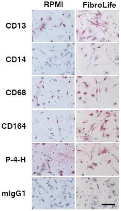
Figure 3. Expression of markers by fibrocytes cultured in RPMI or FibroLife SFM.

PBMC isolated from heparin treated blood were cultured in RPMI- or FibroLife-based serum-free medium for 5 days at 2.5 × 10⁵ cells per ml on 8 well glass slides. Cells were then air-dried, fixed, and stained with antibodies. Cells were counterstained with hematoxylin to identify nuclei. Positive staining was identified by red staining, with nuclei counterstained blue. Bar is 100 μm. The images are representative of three independent experiments.
