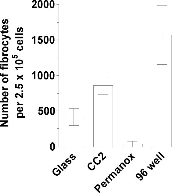
Figure 4. Effect of different substrates on fibrocyte differentiation.
PBMC separated from heparin treated blood were cultured in FibroLife SFM for 5 days at 2.5 × 10⁵ cells per ml on 8-well slides composed of “standard” soda lime glass (glass), positively charged glass (CC2), or plastic (Permanox) substrates. PBMC were similarly cultured in 96 well tissue culture plates (96 well). Results are mean ± SEM (n=3 separate donors).

