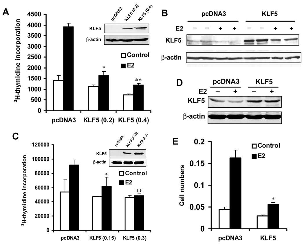
Figure 1. KLF5 inhibits estrogen-induced cell proliferation in ER-positive breast cancer cell lines.
(A–D) DNA synthesis, as indicated by the incorporation of 3H-thymidine (3H-TdR), was used to indicate cell proliferation in both MCF-7 (A) and T-47D (C) breast cancer cell lines. KLF5 expression was restored by transfecting 0.4 µg plasmid DNA including different amounts of pcDNA3-KLF5 and vector control into cells. Expression of KLF5 in transfected cells was confirmed by western blot analysis in both MCF-7 (B) and T-47D (D) cells. E2 treatment was at 1 nM for 20 hours. (E) Cell proliferation was analyzed by colony formation assay in MCF-7 cells. Cells were treated with 1 nM E2 for the last 12 days. Asterisks indicate statistically significant differences in cell growth between cells transfected with pcDNA3 and cells transfected with pcDNA3-KLF5 in the presence of estrogen (p< 0.05).

